


Medical imaging of biomaterials and small animals
The platform offers its expertise to researchers for the selection of the most appropriate small animal imaging method for a given project, as well as a service for developing the associated imaging procedures.
Choice of the most appropriate imaging method (MRI, CT/CT, ultrasound, fluorescence/luminescence, positron emission tomography);
Development of image acquisition protocols appropriate to each type of imaging and experimental procedures;
Standardized experimental protocols, approved by the Laval University animal facilities service;
Housing animals for longitudinal protocols;
Service for the preparation of contrast agents, cell labeling, intravenous injection in-situ (cannulation and injection)
Support for analysis and interpretation of results;
Assistance in writing manuscripts.
The development of more specific imaging protocols can be carried out on request.
All procedures are performed in accordance with the guidelines of the Canadian Council on Animal Care (CCAC) . Each project carried out within the platform must be approved beforehand by the ethics committee in animal experimentation of the research center of the CHU de Québec.
Users must mention, in their publications, the use of the services of the platform in the acknowledgments. The name of each member of the platform substantially involved in a project (excluding routine experiments), must appear as co-author in scientific publications.
Tariff 1 : Academic Université Laval $52/h-research professional + $125/h equipment (MRI, CT, PET)
Rate 2: Academic outside Laval University $52/h-research professional + $200/h equipment (MRI, CT, PET)
Rate 3: Industrial/Institutional non-academic $150/h-research professional + $325/h equipment (MRI, CT, PET)
The development of each new procedure includes up to 3 hours of meeting and preparation time (not billed).
A quote is then developed and submitted to the client, detailing an amount per animal or per image acquisition procedure.
A preliminary experiment could be necessary, because it would allow to better determine the needs or the possibilities of the apparatus according to a precise request.
Contact:
Small animal imaging platform,
CHU de Québec Research Center (CHUL), 2705 boulevard Laurier, Québec (Qc), G1V 4G2
Phone. (418) 525-4444, extension 47115 (Théophraste Lescot), or 42366 (Marc-André Fortin);
Equipment
Magnetic Resonance Imaging (MRI)
Model : Aspect, M2
Description : Medical imaging technique for obtaining 2D or 3D views of the inside of the body in a non-invasive way.
Characteristic : Magnetic field: 1 Tesla (clinical magnetic field); measurements in weighting T1, T2, diffusion; antennae for mice (body, head) and for samples (culture plates, tube boxes, etc.)
Usage :
Statue : Small animal imaging platform - CHU de Québec Research Center (Management: Groupe Fortin)

X-ray computed tomography (CT / CT-X)
Model : General Electric, eXplore Locus 80
Description : Medical imaging technique based on the absorption of X-rays by the tissues to obtain a three-dimensional image of anatomical structures.
Characteristic : Energies 40 – 80 keV; resolutions down to 22 microns. Density image reconstruction software.
Usage :
Statue : Small animal imaging platform - CHU de Québec Research Center (Management: Groupe Fortin)

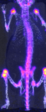
Positron emission tomography (PET)
Model : Imaging research and Technology IR&T, LabPet
Description : Medical imaging technique to obtain a 3D image of the presence of isotope (Beta decay)
Feature : Sub-mm resolution; entire mouse body; static and dynamic measurements
Usage :
Statue : Small animal imaging platform - CHU de Québec Research Center (Management: Groupe Fortin)

Medical image visualization and analysis software
Model : InVicro, VivoQuant (2017)
Description : Medical image visualization and analysis software
Feature :
Usage :
Statue : Small animal imaging platform - CHU de Québec Research Center (Management: Groupe Fortin)
Confocal microscope (Confocal microscopy - CLSM)
Model : Carl Zeiss, LSM800 Axio Observer 7 (2018)
Description : Fluorescence image of many specimens such as opaque samples, gels and hydrogels and tissues
Feature :
Usage :
Statue : Biomaterials characterization platform - CHU de Québec Research Center

Optical coherence tomography ( Optical Coherence Tomography - OCT)
Model : ThorLabs, Telesto (2002)
Description :
Feature :
Usage :
Statue : Biomaterials characterization platform - CHU de Québec Research Center